150 million-year-old pterosaur cold case has finally been solved
Smyth thinks that so few adults show up on the fossil record in this region not only because they were more likely to survive, but also because those that couldn’t were not buried as quickly. Carcasses would float on the water anywhere from days to weeks. As they decomposed, parts would fall to the lagoon bottom. Juveniles were small enough to be swept under and buried quickly by sediments that would preserve them.
Cause of death
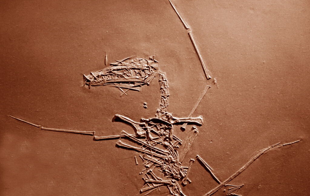
The humerus fractures found in Lucky I and Lucky II were especially significant because forelimb injuries are the most common among existing flying vertebrates. The humerus attaches the wing to the body and bears most flight stress, which makes it more prone to trauma. Most humerus fractures happen in flight as opposed to being the result of a sudden impact with a tree or cliff. And these fractures were the only skeletal trauma seen in any of the juvenile pterosaur specimens from Solnhofen.
Evidence suggesting the injuries to the two fledgling pterosaurs happened before death includes the displacement of bones while they were still in flight (something recognizable from storm deaths of extant birds and bats) and the smooth edges of the break, which happens in life, as opposed to the jagged edges of postmortem breaks. There were also no visible signs of healing.
Storms disproportionately affected flying creatures at Solnhofen, which were often taken down by intense winds. Many of Solnhofen’s fossilized vertebrates were pterosaurs and other winged species such as bird ancestor Arachaeopteryx. Flying invertebrates were also doomed.
Even marine invertebrates and fish were threatened by storm conditions, which churned the lagoons and brought deep waters with higher salt levels and low oxygen to the surface. Anything that sank to the bottom was exceptionally preserved because of these same conditions, which were too harsh for scavengers and paused decomposition. Mud kicked up by the storms also helped with the fossilization process by quickly covering these organisms and providing further protection from the elements.
“The same storm events responsible for the burial of these individuals also transported the pterosaurs into the lagoonal basins and were likely the primary cause of their injury and death,” Smyth concluded.
Although Lucky I and Lucky II were decidedly unlucky, the exquisite preservation of their skeletons that shows how they died has finally allowed researchers to solve a case that went cold for over a hundred thousand years.
Current Biology, 2025. DOI: 10.1016/j.cub.2025.08.006
150 million-year-old pterosaur cold case has finally been solved Read More »