Fungus could be the insecticide of the future
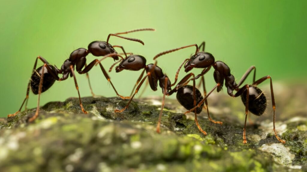
Exterminators keep getting calls for a reason. Wood-devouring insects, such as beetles, termites, and carpenter ants, are constantly chewing through walls or infecting trees and breaking them down. The fight against these insects usually involved noxious insecticides; but now, at least some of them can be eliminated using a certain species of fungus.
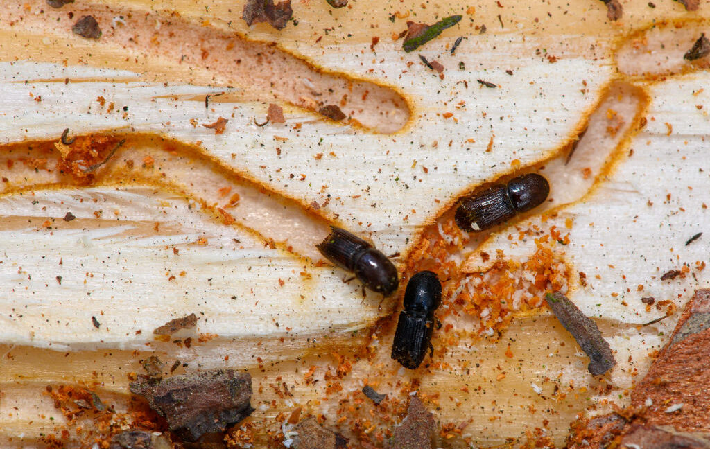
Infestations of bark beetles are the bane of spruce trees. Eurasian spruce bark beetles (Ips typographus) ingest bark high in phenolic compounds, organic molecules that often act as antioxidants and antimicrobials. They protect spruce bark from pathogenic fungi—and the beetles take advantage. Their bodies boost the antimicrobial power of these compounds by turning them into substances that are even more toxic to fungi. This would seem to make the beetles invulnerable to fungi.
There is a way to get past the beetles’ borrowed defenses, though. Led by biochemist Ruo Sun, a team of researchers from the Max Planck Institute for Chemical Ecology in Jena, Germany, found that some strains of the fungus Beauveria bassiana are capable of infecting and killing the pests.
“Insect herbivores have long been known to accumulate plant defense metabolites from their diet as defenses against their own enemies,” she said in a study recently published in PNAS. “However, as shown here for B. bassiana, fungal pathogens are able to circumvent the toxicity of these dietary defenses and cause disease.”
First line of defense
Populations of bark beetles have recently exploded in temperate forests because of climate change. One species they feed on is the Norway spruce (Picea abies), which makes organic phenolic compounds known as stilbenes and flavonoids. Stilbenes are hydrocarbons that function as secondary metabolites for plants, and flavonoids, which are polyphenols, are also secondary plant metabolites that are often antioxidants. The spruce links both classes of compounds with sugars and relies on their antibacterial and antifungal activity.
When metabolized by the beetles, the spruce sugars are removed through hydrolysis, converting them into aglycones that are even more toxic to microscopic invaders. Despite that, some fungi appear to be able to deactivate these compounds. Strains of the fungal insect pathogen B. bassiana have been documented as killing some of these beetles in the wild.
Fungus could be the insecticide of the future Read More »


