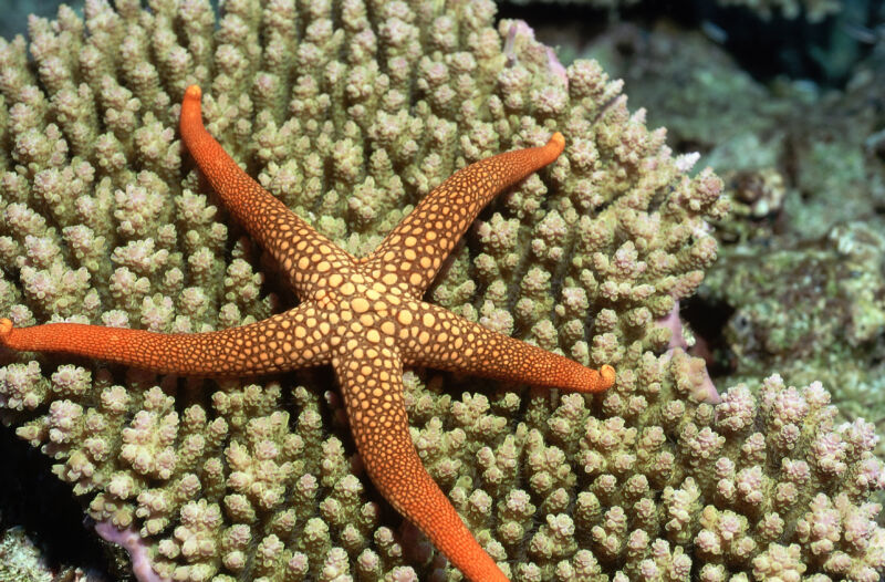
A single peptide helps starfish get rid of a limb when attacked
You can have it —
A signaling molecule that’s so potent injected animals may drop more than one limb.

For many creatures, having a limb caught in a predator’s mouth is usually a death sentence. Not starfish, though—they can detach the limb and leave the predator something to chew on while they crawl away. But how can they pull this off?
Starfish and some other animals (including lizards and salamanders) are capable of autonomy (shedding a limb when attacked). The biology behind this phenomenon in starfish was largely unknown until now. An international team of researchers led by Maurice Elphick, professor of Animal Physiology and Neuroscience at Queen Mary University of London, have found that a neurohormone released by starfish is largely responsible for detaching limbs that end up in a predator’s jaws.
So how does this neurohormone (specifically a neuropeptide) let the starfish get away? When a starfish is under stress from a predatory attack, this hormone is secreted, stimulating a muscle at the base of the animal’s arm that allows the arm to break off.
The researchers confirmed this neuropeptide “acts as an autotomy-promoting factor in starfish and such it is the first neuropeptide to be identified as a regulator of autotomy in animals,” as they said in a study recently published in Current Biology.
Holding on
Elphick’s team studied how the neuropeptide known as ArSK/CCK1 facilitates autonomy in the European Starfish, Asterias rubens. ArSK/CCK1 is already known to inhibit feeding behavior in A. rubens by causing the stomach to contract, and muscle contraction plays a role in limb loss. The researchers found that its ability to trigger contractions goes beyond feeding.
Starfish underwent an experiment that simulated conditions where a predator’s jaw clamped down on one arm. Clamps were placed on one of three sections on a single arm, either on the end, middle, or at the site in the base where autotomy is known to occur, also known as the autotomy plane. The starfish were then suspended by these clamps above a glass bowl of seawater. During the first part of the experiment, the starfish were left to react naturally, but during the second part, they were injected with ArSK/CCK1.
Without the injection, autotomy was seen mostly in animals that had arms that were clamped closest to the autotomy plane. There was not nearly as much of a reaction from starfish when the arms were clamped in the middle or end.
In the second half of the experiment, the clamping used before was combined with an injection of ArSK/CCK1. For comparison, some were injected with the related neuropeptide ArSK/CCK2. A staggering 85 percent of ArSK/CCK1-injected animals that were clamped in the middle of the arm or closer to the autotomy plane exhibited autonomy, and some autotomized additional arms. This only happened in about 27 percent of those injected with ArSK/CCK2.
Letting go
While ArSK/CCK1 proved to be the most effective chemical trigger for autotomy, its activity in the autotomy plane depends on certain aspects of a starfish’s anatomy.
Like all echinoderms, starfish have endoskeletons built of tiny bones, or ossicles, linked by muscles and collagen fibers that allow the animals to change posture and move. Two exclusive features only found in the autotomy plane allow this structure to break. Under the skin of the autotomy plane, there is a region where bundles of collagen fibers are positioned far apart to make breakage easier. The second of these features is a band of muscle close to the region of collagen bundles. Known as the tourniquet muscle, this muscle is responsible for the constriction that allows an arm in danger to fall off.
Analyzing starfish arm tissue while it was undergoing autotomy gave the scientists a new perspective on this process. Right after a starfish has its arm seized by a predator, ArSK/CCK1 tells nerves in the tourniquet muscle to start constricting in the region right by the autonomy plane. While this is happening, the collagen in the body wall in that region softens and breaks, and so do the muscles and ligaments that hold together ossicles. It is now thought that ArSK/CCK1 is also involved in the softening of this tissue that prepares it for breakage.
After starfish autotomize a limb, that limb eventually regenerates. The same happens in other animals that can use autotomy to their advantage (such as lizards, which also grow their tails back). In the future, finding out why some animals have the ability to regenerate may tell us why we either never evolved it or some of our ancestors lost the ability. Elphick acknowledged that there might still be other unidentified factors working together with ArSK/CCK1, but further insight could someday give us a clearer picture of this process.
“Autotomy is a key adaptation for survival that has evolved in several animal taxa,” the research team said in the same study, “[and] the findings of this study provide a seminal insight into the neural mechanisms that control this remarkable biological process,”
Current Biology, 2024. DOI: 10.1016/j.cub.2024.08.003
A single peptide helps starfish get rid of a limb when attacked Read More »


