Human muscle cells come back from space, look aged
Putting some muscle into it —
Astronauts’ muscles atrophy in space, but we can identify the genes involved.

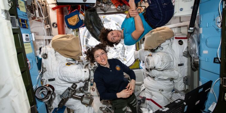
Enlarge / Muscle atrophy is a known hazard of spending time on the International Space Station.
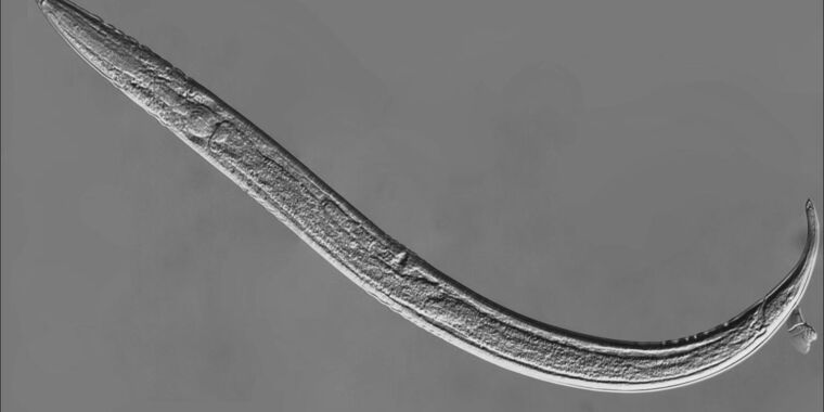
Muscle-on-chip systems are three-dimensional human muscle cell bundles cultured on collagen scaffolds. A Stanford University research team sent some of these systems to the International Space Station to study the muscle atrophy commonly observed in astronauts.
It turns out that space triggers processes in human muscles that eerily resemble something we know very well: getting old. “We learned that microgravity mimics some of the qualities of accelerated aging,” said Ngan F. Huang, an associate professor at Stanford who led the study.
Space-borne bioconstructs
“This work originates from our lab’s expertise in regenerative medicine and tissue engineering. We received funding to do a tissue engineering experiment on the ISS, which really helped us embark on this journey, and became curious how microgravity affects human health,” said Huang. So her team got busy designing the research equipment needed to work onboard the space station. The first step was building the muscle-on-chip systems.
“A lot of what was known about how space affects muscles was gathered through studying the astronauts or studying animals like mice put in microgravity for research purposes,” Huang said. “In some cases, there were also in vitro cultured cells on a Petri dish—something very basic. We wanted to have something more structurally complex.” Her team developed a muscle-on-chip platform in which human myotubes, cells that organize into long parallel bundles that eventually become muscle fibers in a living organism, were grown on collagen scaffolds. The goal was to make the samples emulate real muscles better. But that came with a challenge: keeping them alive on the ISS.
“When we grow cells on Earth, we pour the medium—basically a liquid with nutrients that allow the cells to grow—over the cells, and everything is fine,” Huang said. “But in space, in the absence of gravity, we needed a closed, leak-proof, tightly sealed chamber. The medium was sloshed around in there.”
Oxygen and carbon dioxide levels were maintained with permeable membranes. Changing the medium was a complicated procedure involving syringes and small custom-designed ports. But getting all this gadgetry up and running was worth it in the end.
Genes of atrophy
Huang’s team had two sets of muscle-on-chip systems: one on the ground and one on the ISS. The idea of the study was to compare the genes that were upregulated or downregulated in each sample set. It turned out that many genes associated with aging saw their activity increase in microgravity conditions.
This result was confirmed when the team analyzed the medium that was taken off after the cells had grown in it. “The goal was to identify proteins released by the cells that were associated with microgravity. Among those, the most notable was the GDF15, which is relevant to different diseases, particularly mitochondrial dysfunction or senescence,” said Huang.
Overall, the condition of cells on the ISS was somewhat similar to sarcopenia, an age-related muscle loss disease. “There were some similarities, but also a lot of differences. The reason we didn’t make sarcopenia the main focus of this study is that we know our muscle-on-chip system is a model. It’s mostly muscle cells on a scaffold. It doesn’t have blood vessels or nerves. Comparing that to clinical, real muscle samples is a bit tricky, as it is not comparing apples to apples,” said Huang.
Nevertheless, her team went on to use their ISS muscle-on-chip samples to conduct proof-of-concept drug screening tests. Drugs they tested included those used to treat sarcopenia, among other conditions.
Space drugs
“One of the drugs we tested was the [protein] IGF 1, which is a growth factor naturally found in the body in different tissues, especially in muscles. When there is an injury, IGF 1 activates within a body to initiate muscle regeneration. Also, IGF 1 tend to be declined in aging muscles,” said Huang. The second drug tested was 15-PGDH-i, a relatively new inhibitor of enzymes that hinder the process of muscle regeneration. Used on the muscles-on-chip on the ISS, the drugs partially reduced some of the microgravity-related effects.
“One of the limitations of this work was that on the ISS, the microgravity is also accompanied by other factors, such as ionizing radiation, and it is hard to dissociate one from the other,” said Huang. It’s still unclear if the effects observed in the ISS samples were there due to radiation, the lack of gravity, both, or some additional factor. Huang’s team plans to do similar experiments on Earth in simulated microgravity conditions. “With some of the specialized equipment we recently acquired, it is possible to look at just the effects of microgravity,” Huang said. Those experiments are aimed at testing a wider range of drugs.
“The reason we do this drug screening is to develop drugs that could either be taken preemptively or during the flight to counteract muscle atrophy. It would probably be more feasible, lighter, and cheaper than doing artificial gravity concepts,” Huang said. The most promising candidate drugs selected in these ground experiments will be tested on Huang’s muscle-on-chip systems onboard the ISS in 2025.
Stem Cell Reports, 2024. DOI: 10.1016/j.stemcr.2024.06.010
Human muscle cells come back from space, look aged Read More »


